Mentors Matter: Taking Students to new heights

Published in Dalhousie News on May 11, 2018. full article here.

Two Dalhousie Medicine New Brunswick students are seeing the very real benefits of having a strong mentor. The members of the Class of 2019 have recently had their research results published in a peer-reviewed open access journal, PLOS.

Amy Brown and Intekhab Hossain, both medical students in their third year, have recently co-published their paper with the support and encouragement of their Research in Medicine (RIM) mentor, Dr. Petra Kienesberger.

The paper, titled Lysphosphatidic acid receptor mRNA levels in heart and white adipose tissue are associated with obesity in mice and humans was the result of many hours spent in the laboratory with guidance from Dr. Kienesberger. A cardiovascular researcher at Dalhousie Medicine New Brunswick, Dr. Kienesberger focuses her research on obesity and its impact on heart disease. She is part of a group of researchers and clinical physicians, called IMPART, who are working to provide health care teams with decision-making tools that will help them to assemble the most appropriate teams of specialists to meet patient needs, to provide truly patient-centred care.

Mentorship

Mentorship is a highly valued process of knowledge transfer in many industries, especially research, with many levels of reward for the mentee. Both students credit their mentor, Dr. Kienesberger, for their success with the project and being able to complete the RIM requirements within their first two years, half the allotted time. “She held us to a high standard, which gave us responsibility and ownership over our project,” Amy recalls.

The Research in Medicine program provides medical students with the opportunity to learn the intricacies of research through a mentorship learning experience. This not only includes the technical aspects of conducting research, but also the professional aspects. “Dr. Kienesberger asked for accountability from us not by demanding it, but by practising it herself… This influenced us to also take responsibility for our communication, tasks, deadlines, etc.,” Intekhab explains.

Research

Amy and Intekhab’s research focused on what types of receptors are present in cardiac tissue, the role obesity plays in the presence of these receptors, and if they are correlated to outcomes after cardiac surgery. As medical students they were very excited about this project as it lays the groundwork for potential new treatments and therapies tailored to the needs of the individual patient.

“The idea is that we’ve found the ingredients for someone to develop a recipe that can offset the negative effects of obesity with drugs/treatments and reduce the length of stay in hospitals,” explains Amy. Their research focuses how to influence the metabolic profile of the patient instead of addressing the disease profile. This type of patient-tailored therapy is becoming the focus of translational research.

Amy and Intekhab’s study required them to work with both laboratory and clinical data. For Amy the research project supported her clinical background and her academic background in molecular biology. “It was the perfect marriage of my interests,” Amy says.

For Intekhab, it was her first introduction to laboratory science: “I hadn’t done lab work before and was looking forward to challenging myself, but I wanted to be supported. Dr. Kienesberger was well prepared and great as a mentor.”

Lasting Impact

Both Amy and Intekhab have developed great respect for their mentor, Dr. Petra Kienesberger, the IMPART lab at Dalhousie Medicine New Brunswick and the impact that mentorship can have. Intekhab plans to take what he has learnt from watching Dr. Kienesberger into residency and beyond. “As Petra did with us, at the beginning of my interaction with future students as a mentor, I will get to know their baseline experiences and expectations. This will help me tailor my teaching to maximize their learning experience.”

The impact on both Amy and Intekhab goes far beyond the research accomplishment itself. They have learned the value of mentorship and how to be a mentor. They both had such a positive experience that they highly recommend Dr. Kienesberger to any future RIM students.


DALHOUSIE RECEIVES $5.1 MILLION INVESTMENT IN HEALTH RESEARCH

Published in Dalhousie News on Feb 06, 2018.   

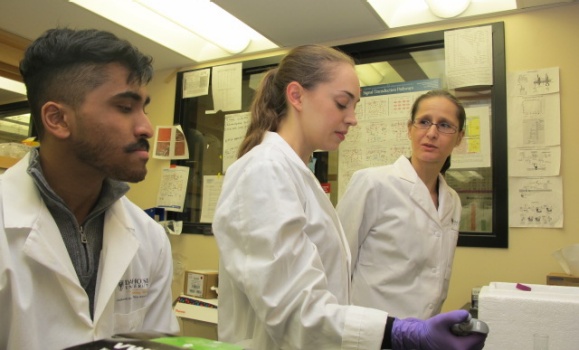
Eight medical scientists from Dalhousie University are the recipients of over $5 million in funding from the Government of Canada for their innovative health research.

Three clinician scientists affiliated with the medical school and Nova Scotia Health Authority also received funding, bringing the total in new health research funding to $7.7 million.

The funding, which is provided by the Canadian Institutes of Health Research (CIHR) project grants, will help Dal’s world-class researchers study a wide range of topics, including: cancer therapy and gene editing; low testosterone and its impact on the heart; pharmaceutical safety and transparency; insulin resistance and heart muscle damage; neurodevelopmental disorders; innate immune memory; more effective breast cancer treatments; and alternative therapies for bacterial infections.

“Our government is fully committed to taking concrete action when it comes to the health and well-being of all Canadians,” said The Honourable Ginette Petitpas Taylor, Minister of Health, in a news release. “This investment will fund research that will lead to new treatments, breakthroughs, and fundamental advances in health science. We are proud of our researchers, and will continue to support them in their efforts to help keep Canadians healthy and continue their research right here at home.”

The Project Grant competition is one of CIHR’s flagship funding programs. Project grants are multi-year grants designed to support researchers at various stages in their careers as they conduct health research and knowledge translation projects that cover the full range of health research topics. Project grant recipients are leaders in their fields and their projects tackle pressing health issues that matter to Canadians, such as cancer, autism, heart disease, and dementia.

“We are proud of the remarkable work being done by our medical researchers, who are improving health and helping us all live healthier lives,” says Alice Aiken, vice-president research for Dalhousie. “We are incredibly grateful for the support they are receiving from CIHR, which enables them to continue leading the way in developing innovative solutions to today’s most pressing health care problems.”

The announcement was made on Wednesday, January 24 at Memorial University of Newfoundland. In total, 512 research projects from across the country will receive $372 million in funding.

Highlights of successfully funded researchers:

Graham Dellaire, Departments of Pathology and Biochemistry & Molecular Biology

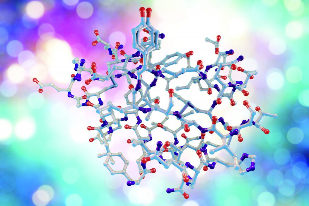
Characterization of HR-Killer1 and identification of small molecules for cancer therapy and enhanced gene editing using CRISPR/Cas9-based DNA repair strategies

Inherited diseases come from mutations in our genes, and genetic mutations are responsible for the development of a variety of cancers. These mutations arise from DNA damage that is repaired incorrectly in our cells. Researchers believe that if you can manipulate DNA repair, you can also enhance DNA damage in cancer cells as a therapy.

In 2013,  it was discovered that DNA in human cells could be efficiently edited using a system from bacteria called CRISPR (Clustered Regularly-Interspaced Short Palindromic Repeats) that works with the cell’s DNA repair machinery. Through the creation of a new test for DNA repair based on CRISPR, Dr. Dellaire and his team have identified a small molecule inhibitor of DNA repair that they call HR-Killer1. With this new funding from CIHR, his team will test the ability of HR-Killer1 to selectively kill cancer cells, and will identify new compounds that enhance CRISPR-based  gene editing for applications in gene therapy.

Susan Howlett, Department of Pharmacology

Impact of low testosterone on cardiac structure and function in aging

Heart disease increases with age in men and women as estrogen and testosterone levels fall. Since a women’s risk of heart diseases goes up after menopause, there has been a lot of interest in the idea the estrogen influences heart disease. However, it is now clear that testosterone decreases with age not only during “manopause” in men, but in women too.

Dr. Howlett and her team are looking at how long term exposure to low testosterone affects how the heart functions in males and females. Their findings from this project will provide a better understanding of the links between testosterone, aging and heart disease. It will also help determine whether testosterone supplementation is good or bad in vulnerable older people with heart diseases.

Matthew Herder, Health Law Institute and Department of Pharmacology

Beyond Transparency in Pharmaceutical Research and Regulation

Making pharmaceutical safety and effectiveness evidence transparent and accessible to physicians, researchers, healthcare payers and patients is an essential but elusive goal.  Although a variety of new laws and policies have been put into place to increase transparency, meaningful improvements have been slow to arrive and changing the real world practices of regulators, clinical researchers  and industry remains a huge challenge.

Professor Herder and his team will work with Health Canada and other key knowledge users to determine the following:
•    How pharmaceutical data can be made more transparent to researchers;
•    what procedural, social and political factors constrain Health Canada’s ability to enforce greater transparency; and,
•    how the regulator can communicate and collaborate with clinical researchers, research institutions and funding agencies to improve pharmaceutical transparency.

Petra Kienesberger, Department of Biochemistry & Molecular Biology

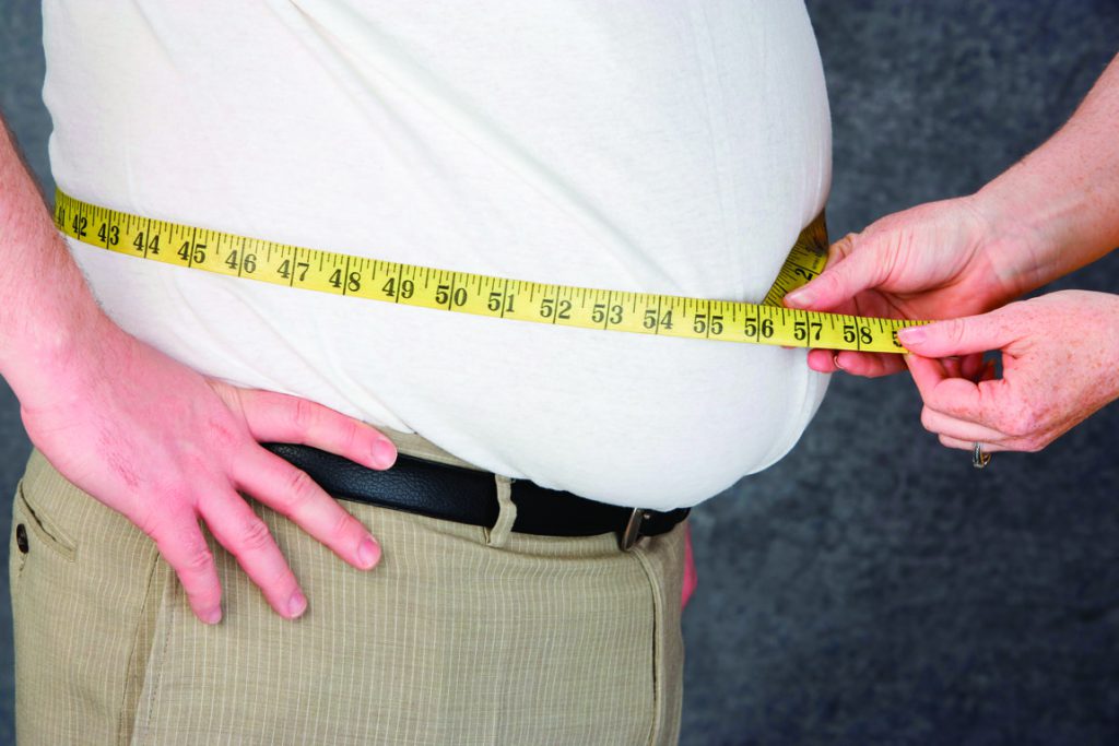
Autotaxin-lysophosphatidic acid signaling in obesity-related heart disease

Obesity is a serious health problem in Canada. Insulin resistance and type 2 diabetes are the main complications of obesity that often damage heart muscle cells, leading to heart muscle weakening and possibly heart failure

Dr. Kienesberger and her team are studying what is happening in heart muscle cells of obese people with insulin resistance or diabetes that leads to heart muscle weakening. They specifically examine the role of bioactive fat molecules in this process, which are released by fat tissue, circulate in the blood stream, and are elevated during obesity and insulin resistance.

Research in the Kienesberger laboratory will help develop new and better ways to treat obesity and diabetes-induced heart muscle weakening, and lower the burden of obesity and diabetes on the Canadian health care system.

Angelo Iulianella, Department of Medical Neuroscience

Molecular regulation of neocortical circuit formation in a model for neurodevelopmental disorders

Neurodevelopmental disorders (NDDs), such as hydrocephaly, lissencephaly, autism spectrum disorder (ASD) and intellectual disability affect up to one in eight children born and represent a significant challenge to the health care system and resources available to Canadian families. NDDs are thought to arise from abnormal brain formation in the fetus, yet the underlying genetic and cellular mechanisms remain largely unknown and treatment options are extremely limited.

Dr. Iulianella and his team have identified a new regulator of neocortical organization that gives several hallmarks of development disorders of the human brain when mutated in mouse models. Their study will help advance our knowledge of the cellular and molecular processes that contribute to the formation of the neocortex and its connections to the regions of the brain important for emotion and cognition. They also hope to identify diagnostic events and therapeutic strategies for addressing developmental disorders of the brain.

Andrew Makrigiannis, Department of Microbiology and Immunology

Understanding Class I MHC Receptor Control of Natural Killer Cell Memory

Adaptive immune memory is one of the most powerful weapons the body has to fight infections. It is essentially how vaccines work: the immune system sees harmful, foreign matter and T and B cells remember it so that the immune response is stronger and faster the next time that target is encountered.

Surprisingly, natural killer cells, which are a cousin to the T cell, can also remember these targets, in a process that is not yet well understood. Dr. Makrigiannis’ research seeks to better understand what Ly49I, a specific natural killer cell protein, does that allows memory in these cells and identify which other proteins and helper cells contribute to natural killer memory.

A whole new field of study in immunology will be opened up by learning how this memory works, and it could also lead to novel cancer vaccines and therapies designed to prevent the recurrence of a dormant cancer.

Kirill Rosen, Department of Biochemistry & Molecular Biology

Molecular hallmarks of breast cancer sensitivity to ErbB2-targeted therapies

Many breast tumors are driven by a protein ErbB2, which they overproduce. These tumors are treated with drugs called ErbB2 inhibitors, but not everyone benefits from them. In addition, these drugs can damage the heart and are costly, so being able to determine who would and would not benefit from them is very important.

Breast tumor cells originate from normal breast cells forming a layer in the breast. Normal cells die when they detach from this layer. ErbB2 blocks the death of cancer cells after they detach, which allows them to form tumors and spread throughout the body. How ErbB2 causes these effects is not well understood.

Dr. Rosen and his team have discovered that ErbB2 triggers signals that cause a loss of a protein, known as Irf6, in tumor cells after they detach from their normal location, and that Irf6 reduces a cellular amount of a cell death-inducing protein known as Perp. Cells lacking Perp do not die outside their normal location.

In this research project, they will examine what signals ErbB2 induces to cause Irf6 and Perp loss in breast tumor cells, and whether this loss allows the cells to form tumors and spread through the body. Some ErbB2-overproducing tumors are treated with ErbB2 inhibitors in an effort to shrink the tumor, which is then surgically removed. The patient further receives these drugs to kill tumor cells that may have stayed in the body.

By testing human breast tumor samples obtained before and after treatment with the drugs, Dr. Rosen will be able to assess whether the increase in Irf6 and Perp levels in the tumor after it is forced to shrink by the drugs predicts whether the patient will benefit from these medicines. Patients that are not expected to benefit from the drugs would be able to avoid them after surgery. They are hoping to identify a new breast cancer mechanism and to develop a method of predicting who will benefit from ErbB2 inhibitors.

Xianping Dong, Department of Physiology and Biophysics

Functional crosstalk between TRPML3 and BK in autophagy induction and pathogen defense

Bacterial infections are the most common medical conditions. They often result in considerable economic and public health burdens and can have a large impact on the quality-of-life of the individuals affected. Although patients suffering from bacterial infections are commonly treated with antibiotics, these treatments can result in the development of antibiotic resistance. For this reason, alternative therapies are needed.

Dr. Dong and his team have discovered that there are two endolysosomal membrane ion channels, called BK and TRPML3, can increase pathogen clearance by increasing autophagy. They also predict that chemicals promoting TRPML3 and BK activity could be used to combat bacterial infection.

Kenneth Rockwood, Division of Geriatric Medicine

Determine how frailty influences the risk and expression of dementia in Alzheimer disease 

Dementia is a clinical syndrome in which impaired memory and thinking interferes with a person’s daily life. The most common cause of dementia is mixed Alzheimer disease (AD) / vascular dementia. AD can be diagnosed clinically when someone is alive and, after death, by brain autopsy. Even so, not everyone who meets the autopsy diagnostic criteria for AD actually has dementia in life. Despite some study, why this discrepancy exists is still unclear.

Dr. Rockwood and his team plan to address it by looking at how overall health affects brain function. To do this, they will measure health problems, which they will quantify with a frailty index. The frailty index counts the number of health problems that an individual has accumulated. Frailty increases the risk for many age-related illnesses (including heart disease, hip fracture and dementia). They want to know if this helps explain why brain autopsy findings alone do not account for the clinical features of dementia.

Dr. Rockwood hypothesizes that the frailer a person is, even though it is more likely that they will have cognitive impairment, this will be less clearly related to their brain autopsy findings. They will use data from two large, community-based, autopsy series in older people (Chicago USA; Cambridge UK). The people in those studies generously agreed to have their memory and thinking tested every 1-2 years and to donate their brains after they had died.

This research team includes physicians who study both dementia in people and AD in brains, as well as scientists skilled in data analysis. They have team members at varying levels of their careers, from PhD student to established professor. Understanding how frailty affects brain function can improve our understanding of how dementia arises, and how it can be treated and even prevented.

John Sapp, Division of Cardiology

Comparing the effectiveness of medication versus cardiac ablation in the treatment of the dangerous arrhythmia, ventricular tachycardia.

The VANISH2 trial is designed to determine the best treatment for people who have life-threatening abnormalities of heart rhythm. Heart attacks leave scars in the heart muscle. The scars can interfere with the normal signaling within the heart that controls the heartbeat. In some cases, the interference can cause a very dangerous abnormal heart rhythm known as ventricular tachycardia (VT). This rhythm is the most common cause of sudden death in Canada.

When patients are at high risk for recurrences of VT, a defibrillator (ICD) can be implanted which can shock the heart back to normal rhythm from a cardiac arrest. These devices are life-saving but do not prevent the abnormal rhythm, they just provide a rescue when it occurs.

In order to prevent dangerous arrhythmias, doctors use strong rhythm control drugs or a procedure called catheter ablation. An ablation is performed by advancing wires through the blood vessels into the heart, using X-rays and other imaging to see where they are and the short circuits within the scar can then be identified and interrupted (ablated). Neither the drugs nor the ablation procedure work perfectly and both carry risk. This trial is designed to determine which treatment is the best.

Patients who have had heart attacks and develop VT will be randomly allocated (50:50) to be treated either with rhythm control drugs or catheter ablation. All patients will receive an implanted defibrillator. We will enroll a total of 366 patients and follow them for at least two years to see which group does the best with respect to recurrent abnormal heart rhythms and survival.

This trial will determine whether the best treatment for VT is heart rhythm drugs or catheter ablation. This evidence will permit more optimal treatment for many thousands of patients worldwide.

Phil Tibbo, Department of Psychiatry

Investigating cannabis effects on brain structure and disease course in early phase psychosis.

Schizophrenia, affecting 1{8617e24ab0b76aabcd10cf8004a7bdc562123dc1ea8adc37299158a7c05423e6} of the population, causes significant burden to individuals, families and society. Fortunately, specialized early phase psychosis (EPP) intervention programs result in positive long term outcomes.

Unfortunately, the high rate of cannabis use in EPP affects these outcomes. Cannabis use results in a higher risk of relapse and severity of symptoms, reductions in individuals following thru with treatment and overall functioning deficits (e.g. work and school) compared to EPP non-cannabis users. Cannabis use can reduce the recovery gains of these young adults. Cannabis, thru its receptors located on the brains white matter (WM) tracts, may be affecting WM development during the critical brain developmental period of young adulthood, resulting in these negative outcomes. Damage to the connections in the brain have themselves been reported in long-term schizophrenia, potentially causing schizophrenia symptoms.

The effects of regular cannabis use, on a potentially already damaged brain in early disease, has not been well studied but we suspect that it too reduces the strength of brain connections but in a different way than schizophrenia.

Dr. Tibbo’s two site study (Halifax, NS; London, Ont), will recruit subjects to four groups: cannabis use disorder (CUD) and non-CUD patients within the first year of entry to the EPP programs, and CUD and non-CUD age and sex matched healthy controls. They will collect detailed information about past and current use of cannabis and clinical measures using established rating scales and methods. They will combine novel brain imaging techniques focusing on WM to generate an understanding of specific WM abnormalities associated with cannabis use in EPP, separate from illness and cannabis effects alone.

This research design will allow a pioneer assessment of the potential negative impact of regular cannabis use longitudinally in this population (baseline and one year later), allowing for more directed education and treatments.


For more information about CIHR’s project grants, visit the CIHR website.

Fuel efficiency and the heart: Dr. Thomas Pulinilkunnil studies role of nutrients in cardiac health and diabetic heart disease

Published in The Philanthropist by DMRF, fall 2015. Read full article here.

Dr. Thomas Pulinilkunnil is learning how and excessive or imbalanced intake of nutrients alters metabolism. leading to metabolic disorders, diabetes and heart disease. At the same time, he’s exploring how age and frailty contribute to these disorders.

“I f we consume excessive amounts of fat, protein or carbs, the process of extracting energy from these fuels is not efficient,” says Dr. Pulinilkunnil, as assistant professor in Dalhousie Medical School’s Department of Biochemistry and Molecular Biology at Dalhousie Medicine New Brunswick (DMNB). “This changes our physiology, stimulating our appetite and cravings for energy-dense foods and leading to storage of fats and carbohydrates.”

In his Saint John lab, Dr. Pulinilkunnil examines how amino acids interact with sugar and fat to drive weight gain, insulin resistance and the accumulation of fat in and around the heart. He’s finding that insulin resistance—a precursor to diabetes which increases with age, overeating and lack of exercise—interferes with the ability of heart-muscle cells to generate energy and rid themselves of cellular waste, leading to a weak and toxin-overloaded heart.

“Diabetes is officially classified as a cardiovascular disease,” notes Dr. Pulinilkunnil, whose interest in diabetes is driven in part by the fact that many of his family members have type 2 diabetes. “Habits are important but genetic factors also come into play… metabolic disorders and diabetes are common is South Asian populations, where diabetes exists in the absence of obesity, unlike North America.”

Using fish strains provided by Dr. Jason Berman in Halifax, Dr. Pulinilkunnil and his team are developing a unique zebrafish model of overfeeding. “We will use this model to study the metabolic mechanisms of weight gain and screen therapeutic agents,” sys Dr. Pulinilkunnil, who’s also working Dr. Petra Kienesberger and Dr. Ansar Hassan to study metabolism in human heart tissues obtained from cardiac surgery patients taking part in Molly Appeal-funded studies in the Maritimes.

Illuminating pathways to the future of health

Published in Scientia, May 17, 2017. .

Non-communicable diseases, such as heart disease, obesity, and diabetes, kill more people worldwide than any other disease. Drs Petra Kienesberger and Thomas Pulinilkunnil at Dalhousie University have dedicated their careers to understanding the molecular underpinnings of these diseases, in the hope of creating a healthier tomorrow.

Heart disease has been the leading cause of death worldwide for over 15 years, killing millions of people each year. Diabetes recently rose to the sixth leading cause of death worldwide, and is projected to move further up the list in the future. These two diseases are often associated and share risk factors, as well as distinct metabolic and molecular signatures. While scientists have a grasp on many factors that are associated with heart disease and diabetes, often the underlying mechanisms are not well understood, presenting a barrier to the development of novel treatments for these conditions. The cornerstone of Dr Kienesberger and Dr Pulinilkunnil’s research program is illuminating the role of cellular energy metabolism in governing outcomes of metabolic heart disease.

Autophagy, Obesity, and the Heart

Diabetes and obesity are often linked with heart disease. In particular, many diabetic and obese patients suffer from a group of conditions known as cardiomyopathy – weakening of the cardiac muscle. The progression of cardiomyopathy often leads to heart failure, thus an understanding of the factors that contribute to and predict cardiomyopathy may help doctors to develop new treatments to reduce and prevent heart disease related deaths in the future. Drs Kienesberger and Pulinilkunnil noted that cardiomyopathy in obese and diabetic patients is often associated with changes in cardiac metabolism that lead to glucolipotoxicity, an accumulation of toxic levels of glucose and fatty acids and their metabolites in the bloodstream and in the cardiac cells. When this occurs, cardiac cells start to struggle with energy regulation and stop disposing and recycling unneeded and damaged proteins, which can ultimately lead to cell death. The Pulinilkunnil laboratory hypothesised that glucolipotoxicity interferes with the cells’ natural waste disposal system and sought to identify and characterise the underlying mechanisms.

Cells get rid of waste, such as used proteins, via intracellular organelles called lysosomes. These tiny vessels engulf unwanted organic materials and digest them through a process known as autophagy. Through autophagy, the cell is able to break down and recycle cellular waste to provide energy for the cell and create fresh building blocks for new proteins and cellular products. When lysosomes are unable to perform autophagy, the cell begins to build up unwanted proteins and waste, which can damage the health of the cell. Lysosome autophagy is controlled by a suite of specialised signalling proteins called transcription factors. One of the most important of these proteins is master regulator transcription factor EB, or TFEB for short. TFEB signals for cells to produce new lysosomes, and coordinates the activity of active lysosomes to keep autophagy running smoothly in the cell. The team speculated that TFEB activity and lysosomal function were disrupted in glucolipotoxic conditions.

To test this, they started with observing the heart tissue of obese and diabetic mice. They found that TFEB was present in lower levels in diseased animals than in healthy controls. Next, they grew rodent cardiac cells in a petri dish, and exposed them to differing levels of glucose and fatty acids, allowing them to observe the response of the cells in detail. The isolated cells exposed to glucolipotoxic conditions expressed lower levels of TFEB and showed impaired lysosomal autophagy. Notably, a particular subset of fatty acids seemed to be most toxic, indicating that this mechanism could be targeted in obesity and diabetes to ameliorate cardiomyopathy. To test whether the rodent results held up in humans, small samples of heart tissue were collected from human patients undergoing heart surgery for various cardiac disorders. Analysis of this tissue revealed that indeed, morbidly obese patients with heart disease had lower levels of TFEB than non-obese patients. These results highlighted the connections between conditions and disease, and revealed impoverished TFEB as a potentially critical player in obesity and diabetes related heart disease.

The Complex Role of Fats in Heart Health and Disease

An interest in the consequences of cardiac cell metabolism and glucolipotoxicity led Dr Kienesberger and colleagues to review the role of lipids in cardiac disease, in an effort to tease out the most damaging fats. Cardiac lipotoxicity, the over-accumulation of specific lipids in heart tissues, often leads to damaged cardiac cells, insulin resistance, metabolic dysfunction, and ultimately cell death. However, there are many types of lipids, and how they interact with cardiac tissue is not well understood, leading to the classifications of ‘good’ and ‘bad’ fats. However, much of the research in this area has focused on a single lipid type, and may be neglecting the bigger picture as to how fat molecules and cardiac cells interact. Dr Kienesberger performed a comprehensive survey of the available research on lipotoxicity and found trends that may indicate a complex role for how lipids interact with cardiac tissue in the development of heart disease.

Heart tissue often prefers to consume fatty acids for energy, but healthy cardiac cells have a more flexible metabolism, consuming both fatty acids and sugars present in the blood stream to keep the heart pumping. Diseased hearts often start to show less metabolic flexibility, relying on fewer energy sources and making cells more vulnerable to impairment. Disease states are often associated with the build-up of fatty plaques in the heart tissue, a situation that occurs when there are more fatty acids available than the heart can metabolise. In studies focused on single types of fats, certain lipids appear to be more toxic than others when they build up in these plaques, while others have been labelled as harmless or ‘good’. Dr Kienesberger’s analysis indicates that the reality is much more complex. Some lipids that appear harmless on their own may have components that are toxic or interact with other molecule to cause damage. Dr Kienesberger argues that more comprehensive research on the networks of these molecular interactions is necessary to form a full picture of the processes that underlie lipotoxicity. However, a consistent finding across all studies is that limiting fatty acid uptake prevents the fatty overaccumulations that contribute to heart disease. Therapeutics that aim to limit fatty acid uptake may help prevent heart damage, regardless of lipid type.

A New Understanding of Chemotherapy Induced Heart Disease

The Pulinilkunnil laboratory further expanded this research to other models of heart failure commonly observed in drug induced cardiomyopathy – specifically that induced by Doxorubicin, a drug used for cancer treatment. Doxorubicin, or DOX, is an effective chemotherapy medication commonly used to treat many different kinds of cancer and notably breast cancer. Chemotherapies are often known for their unpleasant side effects such as hair loss and nausea, but DOX is also known to increase susceptibility to cardiomyopathy specifically in women, through mechanisms that are not fully understood. Prolonged treatment with DOX often leads to heart failure, requiring physicians to balance cancer treatment with heart disease risk. DOX is known to suppress mitochondrial metabolism, alter calcium levels, and interfere with protein break down in many kinds of cells, but its effects on cardiac cell waste processing has only recently been uncovered.

Prior work established that there is a perturbation of lysosomal autophagy activity in the cardiac muscle of DOX treated patients, but it was unclear how DOX might be influencing these processes. The Pulinilkunnil laboratory demonstrated that DOX repressed the expression of TFEB and reduced the activity of cathepsin B – a proteolytic enzyme essential for lysosomal autophagy. Overall, they found that the loss of TFEB reduced the expression of many proteins involved in autophagy, disrupted the normal flux of autophagy activities in the cells, reduced the availability and activity of cathepsin B, and increased the activation of cell death programs.

While DOX appears to disrupt mitochondrial activity, restoration of mitochondrial function has largely failed to prevent the drug’s negative effect on heart health. Dr Pulinilkunnil argues that a component of DOX’s apparent effect on mitochondrial activity is related to its influence on autophagy processes; cardiac cells possess a particularly high number of mitochondria, and when unable to recycle, defective mitochondria quickly become overloaded and unable to function. The Pulinilkunnil laboratory’s careful investigation of the DOX-influence pathways associated with lysosomes and autophagy reveal that the drug interferes with autophagic processes at multiple levels, and the disruption of these pathways is what ultimately results in cardiac damage.

In cardiac tissue, DOX interferes with autophagy by altering lysosome structure and impairing transcription factors that regulate lysosomal activity, including TFEB. This results in a build-up of waste in cardiac cells, creating a toxic environment inside the cell and leading to cell death, causing widespread cardiac tissue damage and eventually resulting in heart failure. Taken together, these effects can lead to widespread cardiac tissue damage, increasing the chances of heart complications following cancer treatment with DOX. Despite this grim result, the team found a silver lining: when TFEB was restored to the cells, many of these effects were reduced and cardiac cells were less likely to undergo programmed cell death. This highlights the potential for TFEB as a research target for preventative cardiac health treatment.

In this regard, the Pulinilkunnil laboratory recently published an in-depth review on this topic, detailing what is known about the molecular relationships between these components of health and illuminating novel pathways through which DOX influences heart health. Dr Pulinilkunnil has grown particularly fascinated with the connections between lysosomal autophagy and cardiomyopathy. Studying the effects of metabolites, drugs, peptides, novel compounds, dietary substances, nutrients on lysosomes and TFEB allows him to reveal the pathways involved in autophagy processes and better appreciate how these entities influence functional outcomes in healthy individuals, with the hope that this increased understanding will lead to advances in our understanding of metabolic heart disease and help discover new therapeutics to boost cardiac health down the line.

Tying Obesity to Disease

For both heart disease and diabetes, a high level of body fat is a risk factor, and excess adipose tissue plays a role in disease development. While these effects are routinely attributed to alterations in metabolism associated with obesity, the hormonal and molecular pathways that contribute to disease progression are not fully known. Autotaxin, or ATX, is a protein produced by adipose cells that is released into the blood stream. Once secreted, ATX produces a fat molecule that triggers important signalling cascades in many cell types of the body. ATX has been implicated in cardiovascular disease and, more recently, obesity and diabetes, but it is currently unclear whether it aggravates or mediates progression of obesity and diabetes. The Kienesberger laboratory sought to better understand the behaviour of this protein and its relationship with blood sugar regulation and metabolism.

The team began by examining ATX blood serum levels in mice that had been raised on either regular chow diet or a diet containing high fat and high sugar (HFHS), and had blood collected either closely following a meal or after a 16 hour fast. Mice on the HFHS diet were overweight and had higher levels of ATX. Mice that had fasted prior to blood collection had lower levels of ATX regardless of body condition. These results confirmed that ATX levels are related to overall fat accumulation, but may fluctuate with meals.

Next, the researchers grew adipose cells in petri dishes to more closely analyse ATX secretion in response to varying doses of glucose and insulin, which controls many of the body’s metabolic processes. They found that fat cells with induced insulin resistance due to their inability to properly respond to insulin showed increased ATX secretion, and this effect could be prevented by adding an insulin sensitiser to the mix. Glucose appeared to have a dose-dependent effect on ATX secretion overall – predictably increasing ATX levels as glucose levels went up. Insulin had a more nuanced effect – a short spike of insulin increased ATX, but chronic exposure to insulin decreased ATX levels. ATX levels appeared to be influenced by both short- and long-term changes in nutritional states, blood sugar levels and insulin.

Making Strides in Human Health

The team’s future research directions will continue to pursue a deeper understanding of human health. With the help of a grant from the Heart and Stroke Foundation of Canada – New Brunswick and the New Brunswick Health Research Foundation, Dr Kienesberger intends to delve deeper into the mysteries of ATX, further teasing apart the relationship between this elusive protein, diabetes, and heart health. Dr Pulinilkunnil recently received a grant from the Canadian Diabetes Association to study the role of TFEB and autophagy in mediating heart disease, with the hopes of developing novel treatments for cardiomyopathy.